

1. INTRODUCTION
Skin and mucosal disorders are pathological events that can have multiple origins, complex development, and combined pharmacological therapies. Some patients may be more susceptible to complications of skin diseases, including difficulty healing, as is the case with diabetics, immunosuppressed patients, and bedridden patients. In this case, the choice of oral and topical medications should be made carefully, focusing on the type of skin lesion and the most appropriate pharmaceutical form for application (Powers et al., 2016; Zheng et al., 2023).
From a tissue pathological point of view, difficulty in repairing skin lesions can generally be caused by a prolonged inflammatory phase of healing, microbial infections such as those caused by bacteria or fungi, diabetes, and slow dermal and epidermal regeneration. Thus, drugs that present epithelial debridement mechanisms, promote collagen synthesis, and have antimicrobial and anti-inflammatory activities are used to accelerate healing or resolve skin conditions (González et al., 2016). In this sense, products of plant origin have wide application for the treatment of skin diseases, as certain species produce metabolites that have synergistic mechanisms, such as flavonoids, which have antioxidant activity and reduce the inflammatory stage of skin regeneration, tannins that reduce the flow of blood in the area and adhere to epithelial proteins to facilitate the closure of wound edges, in addition to terpenes and terpenoids that have antibacterial activity, and mucilages, which have immunostimulant activity, epithelial growth, and promote local hydration (Albuquerque, 2018).
This combination of active metabolites culminates in a synergistic action, enhancing the individual effect of each class of substance, making herbal medicines a highly efficient alternative for the treatment of skin and mucosal diseases (Avci and Gergeroglu, 2019; Majewska and Gendaszewska-Darmach, 2011).
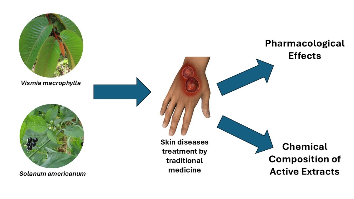
Plants that combine active metabolites with complementary pro-healing mechanisms are found in different biomes around the world, including the Amazon biome. In this sense, among the Amazonian plant resources, there are two species with outstanding ethnomedicinal uses for the treatment of skin diseases and which have recently expanded in terms of scientific knowledge and popular use: Vismia macrophylla Kunth (Hypericaceae) and Solanum americanum Mill. (Solanaceae) (Jara-Herrera, 2023; Mejía-Carhuanca and Rengifo-Salgado, 2000). V. macrophylla is a tree with a stem diameter of 15–20 cm and heights ranging from 10 to 18 m. It is commonly known as “pichiriana” and characterized by secretions of resinous exudates of a reddish-yellow color rich in mono- and sesquiterpenes, which the natives of the Amazon use to treat skin conditions, particularly those caused by fungal infections. However, other species of the genus Vismia are also used by the native people for the treatment of bacterial skin diseases (Jara-Herrera, 2021). On the other hand, S. americanum is a small and widely spreading herb, growing to a height of 150 cm, and although native to South America, it is distributed and adapted on all continents. Because of its very open geolocation, it has various vernacular names, being known in the Amazon by names such as “coconilla,” “Agoema wiwiri,” “Erva-moura,” and “maria-pretinha.” It has a wide variety of ethnomedicinal uses, including vermifuge, as antimalarial and anticancer medication, and also for the treatment of skin conditions such as burns, ulcers, shingles, and “darter.” It is widely used in the Amazon Region for skin ailments, through plaster (Lim et al., 2012; Mejía-Carhuanca and Rengifo-Salgado, 2000).
In spite of the growing scientific interest and the extensive use of these species, there is a dearth of documents on the chemical composition and biological activities pertinent to their use in skin ailments. Therefore, the objective of this review is to comprehensively compile scientific studies related to these topics.
2. RESULTS
2.1. Chemical Information
2.1.1. V. macrophylla
The composition of essential oil (EO) in leaves and fruit was determined in samples from Táchira (Venezuela—February 2013), yielding 1.3% and 5.6% v⁄w, respectively. The main compounds identified were γ-bisabolene (44.4% in leaves and 22.3% in fruits), β-bisabolol (14.9% and 4.4%), trans-Caryophyllene (4.9% and 5.3%), Germacrene D (4.8% and 12.1%), and δ-cadinene (1.4% and 10.7%) (Buitrago et al., 2015) (Table 1). Conversely, a previous study with leaf EO collected in Michelena (Venezuela—June 2010) showed β-caryophyllene (20.1%), Germacrene D (11.6%), β-elemene (7.0%), α-selinene (5.6%), and E-nerolidol (5.0%) as the main constituents, which suggests that the species sensibility is influenced by environmental factors or even the possibility of the existing different chemotypes (Rojas et al., 2011). Recently, another study informed that acetic acid and hexanal are the main compounds in the stem EO of a V. macrophylla specimen found in Northern Costa Rica (Shebitz et al., 2020). Interestingly, the mono- and sesquiterpene compositions from the plant resin are extremely different, according to the study of Jara-Herrera (2023), in which 4-terpineol (36.08%), α-terpineol (21.46%), α-eudesmol (16.89%), α-copaene (8.89%), and camphor (3.99%) are the major substances in the sample collected in Nina Rumi, Peru (2021). Pharmacologically, the terpenoids present in EOs have relevant antimicrobial activity, through nonspecific mechanisms such as the disintegration of the cellular membrane, which helps to prevent wound infection (Albuquerque, 2018).
Table 1
Chemical composition of Vismia macrophylla and Solanum americanum.
| Species | Source | Chemical Composition | Reference |
|---|---|---|---|
| Vismia | EO from leaves (Táchira/Venezuela— | γ-bisabolene (44.4 %), β-bisabolol (14.9 %), trans-Caryophyllene (4.9 %), | (Buitrago et al., 2015) |
| macrophylla | February 2013) | Germacrene D (4.8 %), δ-cadinene (1.4 %) | |
| Kunth. | EO from fruits (Táchira/Venezuela— | γ-bisabolene (22.3 %), Germacrene D (12.1 %), δ-cadinene (10.7 %), trans- | |
| February 2013) | Caryophyllene (5.3 %), β-bisabolol (4.4 %) | ||
| EO from leaves (Michelena/Venezuela— | β-caryophyllene (20.1 %), Germacrene D (11.6 %), β-elemene (7.0 %), | (Rojas et al., 2011) | |
| June 2010) | a-selinene (5.6 %), E-nerolidol (5.0 %) | ||
| EO from stem (Northern Costa Rica) | Acetic acid and hexanal | (Shebitz et al., 2020) | |
| Plant resin (Nina Rumi/Peru—2021) | terpen-4-ol (36,08 %), a-terpineol (21,46 %), a-eudesmol (16,89 %), a-copaene | (Jara-Herrera, 2023) | |
| (8,89 %) and camphor (3.99 %) | |||
| Methanolic, hexanic, and dichloromethanic | triterpenoids, anthrones, small amounts of glycosides, flavones, dehydroflavones, | (Buitrago et al., 2016) | |
| extracts from leaves (Michelena/Venezuela— | and flavonols | ||
| February 2015) | |||
| Young leaves (Soberanía National Park/ | ferruginin A, B, and C, vismin, harunganin | (Hussein et al., 2003) | |
| Panama—April 2002) | |||
| Ethanolic extract from leaves (Brazilian | catechin, osajaxanthone, quercetin, quercitrin, and glucodistylin | (Sanches et al., 2024) | |
| Atlantic Forest) | |||
| Solanum | Leaf crude hydromethanolic extract (Bakin | alkaloids, anthraquinones, cardenolides, saponins, tannins, and flavonoids | (Usman et al., 2018) |
| americanum | Kogi Jalingo/Nigeria) | ||
| Mill. | Leaf aqueous extract (Recife/Brazil—July/ | flavonoids, phenols, and potentially glycoalkaloids | (Bezerra et al., 2025) |
| September 2019) | |||
| Leaves (Igando/Nigeria—5th March 2020) | phenols (64 mg/100 g), alkaloids (59 mg/100 g), flavonoids (47 mg/100 g), | (Nwabiani et al., 2022) | |
| tannins (46 mg/100 g) and reducing sugars (24 mg/100 g) | |||
| Leaves (Zamorano/Honduras) | of protein, lipids, calcium, iron, and vitamin A, and relevant amounts of vitamin C | (Morales, 2008) | |
| and phosphorus | |||
| Fruits (India) | petunidin3-(p-coumaroyl) rhamnosyl glucoside | (Rao, 1978) | |
| leaf extracts (Tübingen/Germany—March/ | diosgenin and tigogenin | (Carle, 1981) | |
| September 1977) | |||
| Aerial parts (Lahore/Pakistan) | α-solamargine (1.96 mg/g), solasonine (3.5 mg/g), α-solanine (3.29 mg/g), | (Mohy-Ud-Din et al., 2010) | |
| solanidine (8.85%), and solasodine (85.67%) | |||
| Aerial parts (Simão Pereira/Brazil) | hydroxybenzoic acid, 3-indolecarboxylic acid, N-trans-p-coumaroyloctopamine, | (Silva et al., 2017) | |
| N-trans-p-feruloyloctopamine, N-trans-p-coumaroyltyramine, and N-trans-p- | |||
| feruloyltyramine. methyl 5-ethyl-4-hydroxy-5-methyl-2-oxotetrahydro-2H- | |||
| pyran-4-carboxylate, loliolide, solameriside A, p-coumaric acid, rhamnetin | |||
| 3-O-β-d-galactoside, and corchorifatty acid B |
Moreover, Buitrago and researchers (2016) report that triterpenoids, anthrones, small amounts of glycosides, flavones, dehydroflavones, and flavonols were found in methanolic hexanic and dichloromethanic extracts of V. macrophylla (Buitrago et al., 2016), whereas at least five active prenylated anthracenes were isolated from the young leaves of V. macrophylla collected in Soberanía National Park (Panama, April 2002): ferruginin A, B, and C, vismin, and harunganin (Hussein et al., 2003). Subsequently, it was identified that catechin, osajaxanthone, quercetin, quercitrin, and glucodistylin are the main flavonoids of its leaf ethanolic extract from leaves (Sanches et al., 2024). In turn, flavonoids and phenolics, such as quercetin and catechin, in addition to anthracenes, also make an important contribution to the healing mechanism, as they are powerful antioxidants and help reduce the time of the inflammatory stage of healing, in addition to some also having antimicrobial activity (Albuquerque, 2018).
2.1.2. S. americanum
The phytochemical results of the leaf crude hydromethanolic extract revealed a major presence of alkaloids, anthraquinones, cardenolides, saponins, tannins, and flavonoids (Usman et al., 2018), while the aqueous extract also showed the presence of flavonoids, phenols, and potentially glycoalkaloids (Bezerra et al., 2025). The quantitative analysis of some metabolites classes in leaves highlighted the notable yield of phenols (64 mg/100 g), alkaloids (59 mg/100 g), flavonoids (47 mg/100 g), tannins (46 mg/100 g), and reducing sugars (24 mg/100 g) (Nwabiani et al., 2022). Moreover, the leaves of S. americanum showed high levels of protein, lipids, calcium, iron, and vitamin A, and relevant amounts of vitamin C and phosphorus (Morales, 2008).
In another study with the aerial parts of S. americanum, hydroxybenzoic acid and 3-indolecarboxylic acid were identified, which are two acids associated with antioxidant properties. Also, four amide alkaloids correlated with α-glucosidase inhibition and radical scavenging reduction: N-trans-p-coumaroyloctopamine, N-trans-p-feruloyloctopamine, N-trans-p-coumaroyltyramine and N-trans-p-feruloyltyramine. Furthermore, the lactones of methyl 5-ethyl-4-hydroxy-5-methyl-2-oxotetrahydro-2 H-pyran-4-carboxylate and loliolide, the steroid solameriside A, the phenolic p-coumaric acid, the flavonoid rhamnetin 3-O-β-d-galactoside, and the corchorifatty acid B were identified (Silva et al., 2017). In previous studies, Rao (1978) identified the anthocyanin petunidin3-(p-coumaroyl) rhamnosyl glucoside in fruits collected in India (Rao, 1978), whereas the alkaloids diosgenin and tigogenin were found in leaf extracts (Carle, 1981). Regarding alkaloids, solasodine was absent in leaf extracts but was detected in unripe fruits, whereas the following steroidal glycolalkaloids were identified and quantified in aerial parts: α-solamargine (1.96 mg/g), solasonine (3.5 mg/g), and α-solanine (3.29 mg/g). Nevertheless, the steroidal aglycones solanidine (8.85%) and solasodine (85.67%) were quantified and expressed as percentages of the total aglycone content (Mohy-Ud-Din et al., 2010).
2.2. Pharmacological information
2.2.1. V. macrophylla
The EO extracted from the fruits of V. macrophylla exhibited antibacterial activity against both gram-positive bacteria (Staphylococcus aureus ATCC 25923 and Enterococcus faecalis ATCC 29212) and gram-negative bacteria (Escherichia coli ATCC 25922), with MIC values ranging from 150 µL/mL to 740 µL/mL. Meanwhile, the oil obtained from the leaves was effective only against the gram-positive bacteria S. aureus (100 µL/mL) and E. faecalis (500 µL/mL). In addition, it displayed anti-yeast activity against Candida albicans CDC-B385 and C. krusei ATCC 6258, both with an MIC of 600 µL/mL (Buitrago et al., 2015) (Table 2). Also, the resin showed moderate antibacterial activity against S. aureus with inhibition haloes of 17.5 and 20 mm in disk and well assays, respectively, and high activity against Staphylococcus epidermidis (22.5 mm and 28 mm) (Jara-Herrera, 2023). On the other hand, the ethanolic extract from leaves, as well as the major flavonoids glucodistylin and quercitrin also displayed antimicrobial activity, showing broad spectrum action against Acinetobacter baumannii, E. coli, Pseudomonas aeruginosa, S. aureus, and E. faecalis (Sanches et al., 2024).
Table 2
Pharmacological activity of Vismia macrophylla and Solanum americanum.
| Species | Source | Pharmacological activity | Reference |
|---|---|---|---|
| Vismia | EO from fruits | Antibacterial against Staphylococcus aureus ATCC 25923, Enterococcus | (Buitrago et al., 2015) |
| macrophylla | faecalis ATCC 29212, Escherichia coli ATCC 25922 | ||
| Kunth. | EO from leaves | Antibacterial against S. aureus and E. faecalis | |
| Antiyeast against Candida albicans CDC-B385 and C. krusei ATCC 6258 | |||
| Plant resin | Antibacterial against S. aureus and S. epidermidis | (Jara-Herrera, 2023) | |
| Leaf ethanolic extract | Antibacterial against Acinetobacter baumannii,E. coli, Pseudomonas | (Sanches et al., 2024) | |
| aeruginosa, S. aureus, and E. faecalis | |||
| Leaf and flower methanolic extracts | Antioxidant through DPPH assay | (Buitrago et al., 2015) | |
| Fractons from leaf methanolic extract | Cytotoxic against cervix epithelial carcinoma (HeLa), breast carcinoma (SKBr3), and prostate carcinoma (PC3) cells | (Rojas et al., 2017) | |
| ferruginin A, B, and C; vismin, | Cytotoxic against MCF-7 (breast), H-640 (lung), and SF-268 (central | (Hussein et al., 2003) | |
| harunganin from young leaves | nervous system) cells | ||
| Solanum | Leaf crude methanolic extract | Antibacterial against Streptococcus pyogenes and P. aeruginosa | (Usman et al., 2018) |
| americanum Mill. | Antifungal against C. albicans | ||
| Antibacterial against Bacillus subtilis, E. coli, P. aeruginosa, and S. aureus | (Valya et al., 2011) | ||
| Antifungal against Aspergillus niger | |||
| Aerial parts decoction | S. aureus, P. aeruginosa, Salmonella typhimurium, and C. albicans | (Sánchez et al., 2003) | |
| Wound healing | |||
| Decoction, methanolic, and ethanolic | Antibacterial against S. aureus and K. pneumoniae | (López et al., 2024) | |
| extracts from leaves | |||
| Leaf alkaloidic extract | Anti-biofilm against S. aureus | (Panazzolo, 2020) | |
| Aqueous, methanolic, and alkaloid | Antioxidant through DPPH assay | ||
| extract from leaves | |||
| Bark methanolic extract | B. cereus, K. pneumoniae, Vibrio cholerae,and S. aureus | (Ayshi and Ritu, 2023) | |
| Antifungal against Penicillium chrysogenum, A. niger, Saccharomyces | |||
| cerevisae,and Mucor hiemalis | |||
| Anti-arthtritic | |||
| Analgesic | |||
| Leaf ethanolic extract | Anti-inflammatory in vivo | (Ukwubile, 2024) |
Regarding the antioxidant capacity measured by the DPPH assay, it was observed that the methanolic extract from leaves and flowers of V. macrophylla displayed a high effect, presenting an IC50 equal to 5.50 µg/mL (Buitrago et al., 2016). Furthermore, different studies evaluated the cytotoxic activity of its extracts. The fractions from the methanolic extract of leaves showed significant inhibition of cervix epithelial carcinoma (HeLa), with values ranging from 6.09 µg/mL to 17.51 µg/mL (hexane and ethyl acetate/butanolic fractions, respectively), breast carcinoma (SKBr3) with an overexpressed gene with values from 12.14 µg/mL to 16.90 µg/mL (water and butanol fractions), and prostate carcinoma (PC3) from 10.91 µg/mL to 17.70 µg/mL (water and dichlorometanic fractions), so that the hexanic and water fractions displayed eight more fold selectively than taxol, for the HeLa and SKBr3 cells assays, respectively (Rojas et al., 2017). Moreover, the five substances identified by Hussein et al. (2003) presented cytotoxic activity on MCF-7 (breast), H-640 (lung), and SF-268 (central nervous system) cells, with their GI50 varying between 3.3 and 7.3 µg/mL (Hussein et al., 2003).
2.2.2. S. americanum
The crude methanolic extract from leaves collected in Bakin Kogi Jalingo (Nigeria) presented antimicrobial activity against Streptococcus pyogenes, P. aeruginosa, and C. albicans evaluated through disk diffusion assay. The most prominent activity was seen on C. albicans, with a zone inhibition of 15 mm. On other hand, S. aureus, Bacillus subtilis, E. coli, and Klebsiella pneumoniae were found to be resistant at all concentrations (Usman et al., 2018). These results were not totally corroborated by Valya et al. (2011). In spite of leaf methanolic extract showing higher antimicrobial activity in comparison to other extracts, it was not active against C. albicans in this study and demonstrated activity on B. subtilis, E. coli, P. aeruginosa, S. aureus, and Aspergillus niger (Valya et al., 2011). This finding can be possibly explained by the difference of some active phytochemicals’ concentration or also by the genetic difference of microorganism strains, which can be exemplified by the work of Sosa Suplapuco (2020). In addition, the aerial plant decoction exhibited antibacterial activity against the microorganisms S. aureus, P. aeruginosa, Salmonella typhimurium, and C. albicans, and accelerated the wound healing process in rabbits and calves, while the aqueous extract from its unripe fruits demonstrated significant contractile uterotonic activity. The authors refer to the presence of saponins and glycoalkaloids as responsible for its pharmacological activity (Sánchez et al., 2003). Besides this, a subsequent study observed minimal vaginal irritability and an absence of erythema and edema in the skin of rabbits using a 30% decoction of the dried leaves (Martínez Guerra et al., 2009). In addition to the methanolic extracts and decoction, the ethanolic extract also demonstrated antibacterial activity against S. aureus and K. pneumoniae, albeit moderately, and low toxicity against Artemia salina (López et al., 2024).
Moreover, the alkaloidic extract from leaves presented anti-biofilm activity on S. aureus (Panazzolo, 2020). The bark methanolic extract also displayed antimicrobial activity by disk diffusion assay, being active against the bacterium B. cereus, K. pneumoniae, Vibrio cholerae, and S. aureus, as well as against the fungus Penicillium chrysogenum, A. niger, Saccharomyces cerevisae, and Mucor hiemalis, so that the best activity was reached on B. cereus (19 mm inhibition using 700 µg/disk) (Ayshi and Ritu, 2023).
S. americanum also showed antioxidant and anti-inflammatory properties. Panazzolo (2020) demonstrated the antioxidant capacity of the aqueous, methanolic, and alkaloid extracts, which in turn presented 50% of antioxidant activity by DPPH assay, when 250 µg/mL was used (Panazollo, 2020). Previously, it was revealed that the substances N-trans-p-coumaroyloctopamine, N-trans-p-feruloyloctopamine, N-trans-p-coumaroyl-tyramine, and N-trans-p-feruloyltyramine demonstrated the best radical scavenging reduction (Silva et al., 2017). Still, the in vivo anti-inflammatory activity of the leaf ethanolic extract, at doses between 300 and 1200 mg/kg, presented a similar effect compared to diclofenac (10 mg/kg), which in turn was measured through paw edema model in Wistar rats, with no evidence of toxicity (Ukwubile, 2024).
Moreover, the methanolic extract of the bark showed a notable 94.59% efficacy in inhibiting arthritis at a concentration of 1000 µg/mL. This level of effectiveness was comparable to sodium diclofenac, which exhibited a rate of 98.19% at the same concentration. The extract also possesses analgesic properties, indicating its potential involvement in both central and peripheral pain pathways. In this study, it was revealed that the administration of the bark extract at a dose of 400 mg/kg produced a significant analgesic effect. Pretreatment with cGMP inhibitor methylene blue further enhanced this effect, increasing the inhibition percentage from 66.75% to 79.70%. In addition, the role of the GABA-benzodiazepine receptor in neuropharmacological activity is of considerable interest, so that, in the open field and hole cross tests, the extract demonstrated a significant impact on motor coordination, showing results comparable to those of diazepam (Ayshi and Ritu, 2023).
3. AUTHOR’S CONSIDERATIONS
The chemical compositions of V. macrophylla and S. americanum were detailed by the mentioned studies, so that V. macrophylla presents relevant EO yield and reveals a remarkable difference regarding the major compound of its composition, varying among γ-bisabolene, β-caryophyllene, and 4-terpineol, which is probably because of factors such as place and time of collection, genetic variability, or soil conditions. In the case of S. americanum, the work of Mohy-Ud-Din et al. (2010) demonstrated that there is a difference in the presence of the alkaloid solasodine according to the organ and the stage of maturation, which would be important for the implementation of quality control of its extracts and maintenance of pharmacological quality.
According to the studies mentioned in this section, the antibacterial and antifungal activity of V. macrophylla and S. americanum, as well as their relevant antioxidant capacity, are proposed mechanisms to explain their use in the treatment of skin diseases, as the reduction of the local microbial load prevents infections at the wound site, whereas the shortening of the inflammatory phase of skin lesions through antioxidant activity decreases the tissue oxidative damage, characterizing a more efficient resolution of scarring (Albuquerque, 2018). The in vivo anti-inflammatory and wound healing assays with S. americanum extracts corroborated this hypothesis, as it was observed that a reduction in paw edema inflammation accelerated healing resolution (Sánchez et al., 2003; Ukwubile, 2024). In addition, the definition of effective doses when using S. americanum extracts, including the study that demonstrated equivalence of the bark methanolic extract to diclofenac in the antiarthritic assay, is another positive factor to take into consideration, and it would help direct future clinical trials. Furthermore, the analgesic effect of S. americanum is also a likely auxiliary event in the case of painful chronic wounds, which may increase medical interest in cases of difficult healing. In addition, the diversity of cytotoxic action of the V. macrophylla extracts may also suggest the development of similar subsequent studies with characteristic cells from skin carcinoma, including melanomas, although the antitumor mechanism has not yet been elucidated.
The antifungal activity exhibited by V. macrophylla may partially account for its traditional use by Amazonian populations in the treatment of fungal skin infections. In addition, the antibacterial effects observed against pathogenic bacteria associated with skin diseases, such as S. aureus and S. epidermidis, support the potential mechanisms underlying the ethnomedicinal application of Vismia species in the treatment of bacterial infections. On the other hand, the ethnomedicinal use of S. americanum in the treatment of burns, ulcers, and herpes zoster may be attributed to the bioactive metabolites identified in the species, whose mechanisms of action align with the pathophysiology of these conditions. For instance, tannins contribute to reduced exudate loss from wounds by promoting vasoconstriction through local protein complexation, while also facilitating faster wound closure by enhancing the approximation of skin edges. Furthermore, the antibacterial properties of tannins, along with those of saponins, flavonoids, and other secondary metabolites, help prevent microbial colonization and infection, thereby supporting the healing process in burns and ulcerative lesions. Regarding the anti-herpetic effect, it is likely primarily attributed to the presence of the major alkaloids found in S. americanum, so that a topical application of a cream containing alkaloids—primarily solamargine, solasodine, and solasonine—extracted from S. americanum demonstrated efficacy in the treatment of Herpes simplex, Herpes zoster, and Herpes genitalis in two clinical studies. In the first study, 46 patients exhibited clinical improvement, with resolution of symptoms occurring within 3–10 days depending on the herpes type, and no adverse effects or recurrences observed over a 9-month follow-up (Chataing et al., 1996). A subsequent study involving 90 patients confirmed similar therapeutic effects, with complete symptom resolution in 5–10 days and 90% of patients remaining recurrence-free after 1 year (Chataing et al., 2001).
S. americanum has a broader geographical distribution, which has led to more extensive investigations into its chemical composition and biological activities compared to V. macrophylla. Consequently, further in-depth studies on V. macrophylla are warranted. These should include the quantification of major constituents in its extracts, as well as qualitative and quantitative analyses of metabolite profiles across different plant organs and collection sites. Moreover, in vivo assays evaluating wound-healing activity should be conducted for this species to provide data that more accurately reflect their potential therapeutic applications in humans. Another factor to discuss is the discrepancies in antimicrobial activity observed in different studies, offering possible explanations such as differences in phytochemical concentrations or genetic variations in microorganism strains.
Finally, studies on the development of efficient topical formulations with extracts from both species, as well as clinical trials in humans, are considered steps to be followed, aiming at the optimal use of these resources for the treatment of skin conditions, as well as the chemical standardization of their raw materials, which ensures the quality, safety, and efficiency of the products to be used.
4. CONCLUSION
The chemical and pharmacological studies with V. macrophylla and S. americanum demonstrate the potential of both species in the treatment of skin conditions, because of their extensive antimicrobial activities, including against microorganisms that cause skin infections, and antioxidant activity, which reduces the inflammatory stage of healing. In addition, S. americanum also showed anti-inflammatory, analgesic, and healing activity in vivo, while V. macrophylla also demonstrates extensive cytotoxic activity. Therefore, the two species considered in this review are potential active ingredients for topical formulations to be used for skin conditions, and further studies should be carried out in order to critically measure the clinical application and stability of the formulations.
ACKNOWLEDGEMENT
The authors thank to Universidad Nacional de la Amazonia Peruana for technical support in data access.
AUTHOR CONTRIBUTIONS
Cleto Jara-Herrera was in charge of research concept and design, and collection and/or assembly of data; Ricardo Diego D.G. de Albuquerque did data analysis and interpretation, and writing of the article; Frank Romel León-Vargas was concerned with critical revision of the article; Yessenia Vanessa Sherrezade Ramos-Rivas did collection and/or assembly of data; Rosa Isabel Souza-Nájar did critical revision of the article; Carmen Patrícia Cerdeña del Águila gave the final approval of the article.
CONFLICT OF INTEREST
Given his role as Associate Editor, Ricardo Diego D.G. de Albuquerque has not been involved and has no access to information regarding the peer review of this article. Full responsibility for the editorial process for this article was delegated to Editor-in-Chief Kannan RR Rengasamy. The authors declare no conflict of interest.