

1. INTRODUCTION
The use of medicinal plants in traditional healthcare systems has long been a cornerstone of human civilization, providing therapeutic remedies for a wide range of ailments. Among them, sexual dysfunction remains a major worldwide health concern influencing psychological as well as physical well-being. Plant-based aphrodisiacs that offer safer, all-natural alternatives are attracting more attention as rising concern about the adverse effects and contraindications associated with traditional treatments, including phosphodiesterase-5 inhibitors (e.g., sildenafil), grows. Through many pathways—including hormone regulation, vasodilation, and antioxidant activity—herbal medicines have shown promise in improving libido, erectile function, and reproductive health (Rao et al., 2015; Sathyanarayana Rao et al., 2015).
Native to tropical South America, M. jalapa L., often known as the “Four o’clock flower,” is a member of the Nyctaginaceae family and is now somewhat extensively scattered across India, Southeast Asia, and Africa. Various parts of the plant especially the root have long been used historically to treat gastrointestinal problems, inflammation, and
most importantly as an aphrodisiac. Ethnobotanical research documents its usage in improving male sexual performance and reproduction in rural and tribal populations. Despite its longstanding repute, scientific confirmation of its aphrodisiac qualities using contemporary analytical and computational instruments is still rare (Defeudis et al., 2022; Lew-Starowicz and Czajkowska, 2022; Ljungman et al., 2020).
Identification of the bioactive molecules causing pharmacological action of a plant depends on phytochemical profiling. Methods such liquid chromatography-mass spectrometry (LC-MS) and gas chromatography-mass spectrometry (GC-MS) allow exact characterization of complicated plant extracts (Ammar et al., 2018; Uddin et al., 2012). Many of the flavonoids, alkaloids, terpenoids, and phenolic acids found in past research on M. jalapa have been connected to enhanced sexual health in other medicinal plants. Complete phytochemical analysis of the root extract may provide the molecular foundation for its conventional aphrodisiac action (Alam et al., 2016; Katiyar et al., 2016).
By mapping relationships among phytoconstituents, protein targets, and biological pathways, fast-developing field of network pharmacology enables systems-level knowledge of herbal therapy. This method respects the complexity of plant-based treatments and fits the multicomponent nature of herbal extracts. Network pharmacology can predict how the components in M. jalapa alter important pathways involved in sexual function, including nitric oxide signalling, androgen receptor activation, and hormone control by combining phytochemical data with databases like STITCH, SwissTargetPrediction, and KEGG (Ekbbal et al., 2023; Gaurav et al., 2022, 2023).
Furthermore, the present study holds significant value as it bridges traditional ethnomedicinal knowledge of M. jalapa with modern pharmacological validation. By employing an integrated approach—phytochemical profiling, antioxidant assays, thin-layer chromatography (TLC), LC-MS metabolite identification, and network pharmacology—the research not only confirms the presence of bioactive compounds with aphrodisiac relevance but also elucidates their mechanistic pathways. The novelty lies in the comprehensive mapping of identified metabolites to key reproductive and hormonal genes, highlighting their potential role in sexual health and vascular function. This work provides the first multidisciplinary evidence-based framework for developing M. jalapa root extract as a safe, natural therapeutic for sexual dysfunction.
2. MATERIALS AND METHODS
2.1. Glassware, reagent, and software
Glassware such as beakers, conical flasks, measuring cylinders, pipettes, and volumetric flasks is required for precise experimental procedures. Analytical-grade reagents like solvents (ethanol/methanol), standards (curcumin), 2,2-diphenyl-1-picrylhydrazyl (DPPH), and reagents for phytochemical assays are necessary. High-performance thin-layer chromatography (HPTLC) or spectrophotometry was used to assist in quantification. Software tools such as Cytoscape (Version: 3.9.1), STRING, and SwissTargetPrediction are critical for network pharmacology analysis, pathway exploration, and target identification.
2.2. Preparation of extract
To prepare a methanolic extract of M. jalapa (50) using the sonication method, the dried root is weighed accurately and soaked in methanol in a 1:10 ratio (w/v). The mixture is transferred to a sonication bath or probe sonicator and subjected to ultrasonic waves for 30-60 minutes at a controlled temperature (25-30°C). Sonication facilitates the release of bioactive compounds by disrupting the plant cell walls. After sonication, the mixture is filtered using Whatman filter paper, and the filtrate is concentrated under reduced pressure using a rotary evaporator (Attri et al., 2023). The concentrated extract is stored in an airtight container for further analysis (Gaurav et al., 2022).
2.3. In vitro antioxidant analysis
2.3.1. DPPH antioxidant activity
The in vitro antioxidant activity of M. jalapa extract can be assessed using DPPH (2,2-diphenyl-1-picrylhydrazyl) assay. A methanolic DPPH solution (0.1 mM) is prepared and 180µl mixed with 20 µl of different concentration (62.5-1000 µg/mL). The mixture is incubated in the dark at room temperature for 30 minutes. The reduction of DPPH radicals is measured spectrophotometrically at 517 nm. The decrease in absorbance indicates the free radical scavenging activity. The percentage inhibition is calculated using the formula: % inhibition = [(Control absorbance − Sample absorbance) / Control absorbance] × 100. The IC50 value, indicating the concentration required to inhibit 50% of DPPH, is determined (Gaurav et al., 2020).
2.3.2. ABTS antioxidant activity
The in vitro antioxidant activity of M. jalapa extract was evaluated using the ABTS (2,2’-azino-bis-3-ethylbenzothiazoline-6-sulfonic acid) radical cation decolorization assay. To prepare the ABTS•+ solution, 7 mM ABTS was mixed with 2.45 mM potassium persulfate and incubated in the dark at room temperature for 12-16 hours. This solution was diluted with methanol to an absorbance of 0.70 ± 0.02 at 734 nm. Then, 180 µL of the ABTS•+ solution was mixed with 20 µL of M. jalapa extract at varying concentrations (62.5-1000 µg/mL). The mixture was incubated at room temperature for 30 minutes in the dark. Absorbance was recorded at 734 nm using a spectrophotometer. The percentage of ABTS radical scavenging activity was calculated as
The IC50 value, indicating the concentration required to inhibit 50% of radicals, was determined.
2.4. HPTLC analysis
The HPTLC method for analyzing M. jalapa extract involves applying methanolic extract and standard curcumin solutions onto a silica gel 60 F254 HPTLC plate using an applicator. The plate is developed in a presaturated twin-trough chamber with a mobile phase in an optimized ratio (toluene:ethyl acetate:formic acid, 4:6:1 v/v). After development, the plate is air-dried and visualized under UV light (254 and 366 nm). The bands are identified based on their color and Rf value. The method ensures precise determination of compound bands in the extract (Gaurav et al., 2020).
2.5. LC-MS analysis
For the LC-MS analysis, the root extract of M. jalapa L. was prepared at a concentration of 1 mg/mL using distilled water as the solvent. The prepared solution was thoroughly mixed and then filtered using a polyvinylidene fluoride syringe filter to ensure the removal of particulate matter and prevent clogging of the LC-MS instrument. The filtered sample was subjected to analysis using liquid chromatography-mass spectrometry (LC-MS) under isocratic elution conditions on C18 column 100 mm × 4.6 mm i.d. 5µps. The mobile phase consisted of acetonitrile and 0.1% orthophosphoric acid in water mixed in a 30:60 (v/v) ratio. This solvent system was chosen to provide optimal separation and ionization of the phytoconstituents present in the extract. The flow rate was maintained at 1 mL/min throughout the run to ensure steady and reproducible chromatographic separation. The LC-MS system was calibrated, and standard operating parameters were optimized to detect a broad range of compounds with varying polarities and molecular weights. The resulting chromatographic data and mass spectra were analyzed to identify and characterize the major phytochemicals based on their retention times, molecular ions, and fragmentation patterns, thus contributing to the phytochemical profiling of M. jalapa L. root for further pharmacological correlation (Atrooz et al., 2024; Gautam et al., 2023; Salar et al., 2023).
2.6. Network pharmacology
The network pharmacology methodology for exploring M. jalapa therapeutic potential in aphrodisiac activity involved a systematic and integrative approach to identify bioactive compounds, molecular targets, and associated pathways. In this analysis, the bioactive compounds of M. jalapa were identified through LC-MS analysis. The compounds were then screened for potential therapeutic targets based on the different hundreds of genes (Screened from gene card: https://www.genecards.org/) involved in aphrodisiac function. The interaction between compounds and genes was then predicted for network association (compounds and protein interaction network) to determine the protein and compound interaction. The screening was performed through the Cytoscape software (3.9.1). During analysis, only prominent proteins or genes were screened for gene ontology analysis, while genes that showed no interaction with the compounds were be removed from the final network (Gaurav et al., 2020, 2023).
2.7. Gene ontology DisGeNET analysis
Gene Ontology (GO) and DisGeNET analysis were performed using Metascape to identify key biological pathways and mechanisms involved. This integrative approach was elucidated as multitarget, multipathway mechanism of M. jalapa in aphrodisiac potential, providing insights into its therapeutic action and potential for novel drug discovery. The genes that showed prominent interaction were mapped during GO analysis (Ekbbal et al., 2023; Gaurav, 2022).
3. RESULTS AND DISCUSSION
The methanolic extraction of M. jalapa roots was successfully carried out using the sonication-assisted extraction method. The air-dried and powdered plant material (50 g) was subjected to ultrasonic-assisted extraction with methanol as the solvent. The sonication process was conducted at room temperature for 30 minutes to enhance solvent penetration and improve extraction efficiency. After filtration, the extract was concentrated under reduced pressure using a rotary evaporator and then dried to obtain a semisolid residue. The extractive yield was calculated based on the initial weight of the plant material and the final weight of the dried extract. The methanolic extract yielded approximately 6.42 g, corresponding to an extractive yield of 12.835% w/w. This result indicates a moderately high recovery of phytochemicals using the sonication method. The dark brown, viscous extract obtained was stored at 4°C for further phytochemical screening and biological activity analysis, including antioxidant and anti-inflammatory assays.
3.1. In vitro antioxidant activity
3.1.1. DPPH antioxidant activity
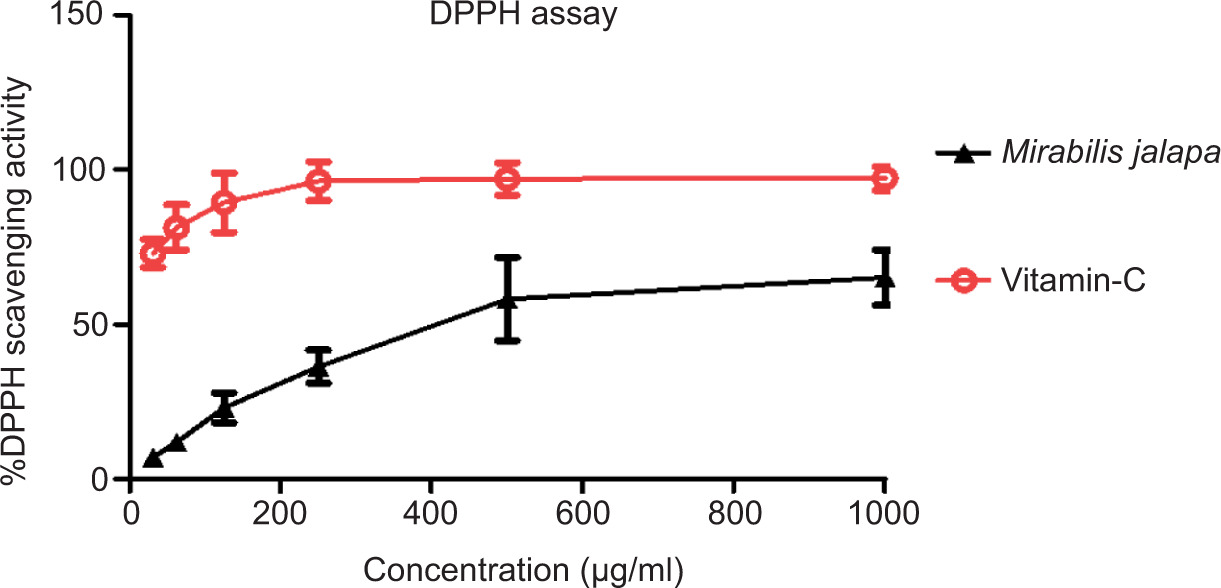
The antioxidant activity of M. jalapa root extract was evaluated using the DPPH (2,2-diphenyl-1-picrylhydrazyl) radical scavenging assay. The graphical data provided shows a dose-dependent increase in the percentage inhibition of DPPH radicals with increasing concentrations of the methanolic extract of M. jalapa. This trend is indicative of the free radical scavenging ability of the extract, suggesting its potential as a natural antioxidant.
In the graph, the x-axis represents the logarithmic concentration of the extract (log [Conc]) in µg/mL, while the y-axis shows the percentage of DPPH radical inhibition. The plotted data points show a clear sigmoid (S-shaped) curve, which is characteristic of dose-response relationships in biological systems. The error bars represent standard deviations, indicating experimental reproducibility and statistical reliability.
At lower concentrations (62.5-250 µg/mL), a sharp increase in DPPH inhibition was observed. For instance, at 62.5 µg/mL, the inhibition was approximately 20-25%, and it increased to around 60-70% at 250 µg/mL. The activity plateaued between 500 and 1000 µg/mL, reaching above 85% inhibition, indicating saturation of the scavenging capacity at higher concentrations. This plateau suggests that the DPPH radicals in the solution were effectively neutralized and that increasing the extract concentration further had a minimal effect on inhibition percentage.
The IC50 value, the concentration required to inhibit 50% of DPPH radicals—was estimated to fall between 150 and 250 µg/mL, based on the curve’s midpoint. This IC50 value reflects the potency of the extract and is commonly used to compare antioxidant strength. A lower IC50 indicates higher antioxidant activity. Compared to synthetic antioxidants like ascorbic acid (which generally have IC50 values in the range of 10-50 µg/mL), the M. jalapa extract exhibits moderate antioxidant activity, which is significant for a crude plant extract.
These findings correlate with previous studies that have identified several antioxidant phytoconstituents in M. jalapa, including flavonoids, phenolic acids, alkaloids, and glycosides. Flavonoids, in particular, are known for their electron-donating properties, allowing them to reduce the DPPH radical into a stable diamagnetic molecule. The high scavenging activity observed in the extract is likely attributable to the presence of such compounds, which interact with free radicals by donating hydrogen atoms or electrons.
Furthermore, the sonication-assisted extraction method used to obtain the methanolic extract enhances the release of phytochemicals by disrupting cell walls and increasing solvent penetration. This method has been shown to improve the efficiency and yield of bioactive compounds compared to conventional maceration or Soxhlet extraction. The extractive yield of 12.835% supports the effective recovery of antioxidant constituents, which likely contributed to the strong DPPH scavenging activity observed.
The stability of the scavenging activity at higher concentrations also suggests a saturation point beyond which the antioxidant potential does not significantly increase. This is important from a pharmacological perspective, as it helps determine the optimal dosage range for therapeutic efficacy without unnecessary overdosing.
Reactive oxygen species and free radicals are known to play a significant role in the pathogenesis of various diseases, including cancer, cardiovascular disorders, diabetes, and neurodegenerative conditions. The neutralization of these free radicals through antioxidant compounds is a crucial strategy in preventing and managing oxidative stress-related conditions. In this context, the moderate but significant antioxidant activity of M. jalapa presents a promising natural therapeutic option. The use of natural antioxidants is increasingly favored over synthetic ones due to safety concerns, potential toxicity, and side effects associated with long-term synthetic antioxidant use. Hence, plant-based extracts with antioxidant properties, such as M. jalapa, are valuable candidates for incorporation into pharmaceutical and nutraceutical formulations.
Moreover, the antioxidant potential of M. jalapa could complement its previously documented pharmacological activities, including anti-inflammatory, antimicrobial, wound healing, and hepatoprotective effects. The oxidative stress pathway is intimately linked to inflammation, making antioxidants potential adjuvants in anti-inflammatory therapies. While the current DPPH assay provides valuable insights into the antioxidant potential of the extract, it is limited to a single mechanism of radical scavenging and does not fully capture the complexity of oxidative processes in biological systems. Other in vitro antioxidant assays, such as ABTS, ferric reducing antioxidant power (FRAP), and oxygen radical absorbance capacity (ORAC), should be performed to obtain a more comprehensive antioxidant profile (Gaurav et al., 2020, 2023; Parveen et al., 2020). Therefore, the DPPH assay results demonstrate that the methanolic extract of M. jalapa exhibits significant dose-dependent antioxidant activity. With an extractive yield of 12.835% and an IC50 estimated between 150 and 250 µg/ml, the extract shows promising radical scavenging potential attributed to its rich phytochemical content. These findings support the traditional medicinal use of M. jalapa and lay a strong foundation for future pharmacological and phytochemical research.
3.1.2. ABTS antioxidant activity
The antioxidant capacity of M. jalapa methanolic root extract was assessed through the ABTS [2,2'-azino-bis(3-ethylbenzothiazoline-6-sulfonic acid)] radical scavenging assay. The plotted graph illustrates the dose-dependent inhibition of ABTS radicals across a range of extract concentrations, thereby indicating the potential antioxidant efficacy of the extract. The x-axis represents the logarithmic concentration of the extract in µg/mL, and the y-axis denotes the percentage inhibition of the ABTS radical.
From the graph, a sigmoidal dose-response curve is evident. As the concentration of the extract increased from 62.5 to 1000 µg/mL, the percentage of ABTS radical inhibition increased accordingly. At lower concentrations (62.5 and 125 µg/mL), inhibition values were modest approximately 20-30%. However, between 250 and 500 µg/mL, a rapid increase in scavenging activity was observed, reaching above 70% inhibition. At concentrations from 500 to 1000 µg/mL, the inhibition curve plateaus near the 85-90% range, suggesting a saturation point where most ABTS radicals in the solution had been neutralized. This behavior is typical of antioxidant activity, where the scavenging effect increases with concentration but reaches a maximum limit due to the finite number of radicals available in the solution. The extract thus demonstrated strong free radical-neutralizing activity, particularly evident from the steep slope of the curve between 125 and 500 µg/mL.
The IC50 value, representing the concentration at which 50% inhibition of ABTS radicals occurred, was found between 200 and 250 µg/mL. This value provides a quantitative estimate of the antioxidant potency of the extract. While this IC50 value may be higher than that of standard antioxidants such as ascorbic acid or trolox (which often exhibit IC50 values in the range of 10-50 µg/mL), it is highly encouraging for a crude plant extract. The result reflects the presence of natural antioxidant compounds with significant biological potential. The error bars associated with each data point indicate a relatively small standard deviation, confirming the reproducibility and statistical reliability of the results. The consistency between the replicates strengthens the evidence supporting the potent antioxidant activity of the M. jalapa extract.
Figure 1
DPPH radical scavenging activity of Mirabilis jalapa methanolic extract. The figure shows the dose-dependent DPPH free radical scavenging activity of M. jalapa root extract. Increasing concentrations (62.5-1000 µg/mL) demonstrate a steady rise in inhibition percentage, indicating potent antioxidant potential. The results represent mean ± SD of three independent experiments.
These findings can be correlated with the phytochemical profile of M. jalapa, which includes flavonoids, phenolic compounds, alkaloids, and betalains. Flavonoids and phenolics are well-established antioxidants capable of donating hydrogen or electrons to neutralize free radicals like ABTS•+. In particular, the hydroxyl groups present in flavonoid structures enhance their antioxidant action through metal chelation and free radical neutralization mechanisms. The substantial inhibition of ABTS radicals suggests that these bioactive constituents are present in sufficient quantities in the methanolic extract to exert significant activity. It is noteworthy that the ABTS assay allows the evaluation of both hydrophilic and lipophilic antioxidant compounds, which gives it an advantage over DPPH assay which is more suitable for hydrophobic systems. Therefore, the observed antioxidant activity in this assay might better reflect the total antioxidant capacity of the extract, especially since methanol, as a polar solvent, is efficient in extracting both phenolic and flavonoid compounds.
In addition to their free radical scavenging activity, antioxidants like those found in M. jalapa have been linked to anti-inflammatory, antimicrobial, and antiaging properties. Oxidative stress, resulting from an imbalance between free radicals and antioxidants in the body, plays a crucial role in the pathophysiology of chronic diseases such as cancer, cardiovascular disorders, neurodegenerative diseases, and diabetes. Hence, antioxidants derived from natural sources are of growing interest for the development of functional foods and therapeutic agents. The ABTS assay involves the generation of the blue-green ABTS radical cation (ABTS•+), which absorbs at 734 nm. When antioxidants in the sample reduce this radical, a decrease in absorbance is observed. The extent of this reduction is proportional to the antioxidant capacity of the test sample. In this experiment, the clear dose-dependent reduction in absorbance demonstrates the efficient interaction between the plant extract’s bioactive compounds and the ABTS radicals. The near-saturation of inhibition values at higher concentrations indicates that the antioxidant compounds have effectively scavenged most of the available radicals, and adding more extract does not significantly improve the result. This plateau is crucial in identifying the optimal range of extract concentration that provides maximal activity without unnecessary excess.
Comparing this ABTS-based activity with earlier DPPH assay results, both assays revealed a consistent antioxidant profile for M. jalapa. However, the IC50 value from the ABTS assay appeared slightly higher than that from the DPPH assay, possibly due to differences in solubility and assay conditions. These differences highlight the importance of using multiple assays to evaluate antioxidant potential comprehensively. To further confirm and elucidate the antioxidant mechanisms, additional in vitro assays such as FRAP, ORAC, and nitric oxide scavenging should be considered. Moreover, in vivo studies are essential to validate the extract’s efficacy under physiological conditions. Chromatographic techniques like HPLC and LC-MS should also be employed to identify and quantify the key antioxidant compounds. The ABTS radical scavenging assay demonstrates that the methanolic extract of M. jalapa roots possesses significant antioxidant activity in a concentration-dependent manner. With inhibition values approaching 90% at higher concentrations and an IC50 value between 200 and 250 µg/mL, the extract exhibits potent free radical neutralization potential. The presence of flavonoids and phenolic compounds likely contributes to this effect. These findings support the traditional medicinal use of M. jalapa and encourage its continued exploration as a natural source of antioxidants for pharmaceutical and nutraceutical applications.
3.2. HPTLC analysis
Thin-layer chromatography (TLC) is a rapid and effective analytical method for the preliminary identification of phytoconstituents in plant extracts. In this study, the methanolic extract of M. jalapa was subjected to TLC profiling to identify the presence of bioactive compounds using different visualization techniques: shortwave UV (254 nm), longwave UV (365 nm), and iodine vapor (visible light). The TLC plate image consists of two tracks labeled (A) and (B). Track A represents the M. jalapa extract, and track B is the standard reference or blank. The mobile phase used was likely a nonpolar to moderately polar solvent system, suitable for the separation of phytochemicals such as alkaloids, flavonoids, terpenoids, and phenolic compounds. Under 254 nm UV light, track A displays a few dark quenching zones against a fluorescent green background, indicating the presence of UV-absorbing compounds. At least two major spots were observed at different positions. The presence of such spots under shortwave UV typically points to aromatic or conjugated systems, likely flavonoids or phenolics. Track B (control or solvent blank) showed no visible spots, confirming the specificity of the extract.
Figure 2
This graph illustrates the ABTS radical inhibition by Mirabilis jalapa methanolic extract across various concentrations. A concentration-dependent increase in scavenging activity is evident, plateauing at higher doses. The data, expressed as mean ± SD, confirm the extract’s strong antioxidant efficacy through ABTS assay.

Under 365 nm UV light, the TLC plate revealed blue and faint green fluorescent bands in track A, suggesting the presence of compounds such as flavonoids, coumarins, or essential oils that fluoresce under long-wave UV. Fluorescence is generally an indicator of conjugated double bonds and certain substituted phenols, implying the presence of potent antioxidant components in the extract. Again, track B showed no fluorescence.
Exposure to iodine vapors results in yellowish-brown spots on the TLC plate, which indicates the presence of nonpolar compounds such as steroids, terpenoids, and certain lipophilic alkaloids. The iodine-reactive spots were distinct and dense in track A, confirming the presence of such classes of compounds in M. jalapa extract. No visible spot was observed in track B.
3.3. Rf value determination
Rf (Retention factor) is calculated as:
The TLC analysis of M. jalapa methanolic extract reveals a diverse phytochemical profile. The presence of multiple bands across different Rf values under various detection conditions highlights the complexity and richness of the extract in secondary metabolites. Under UV 254 nm, the detection of distinct spots indicates the presence of compounds with aromatic structures such as flavonoids and phenolics, which are known for their antioxidant, anti-inflammatory, and antimicrobial properties. These compounds usually absorb in the UV region due to their conjugated π-electron systems. Under UV 365 nm, several fluorescent spots were observed, supporting the presence of polyphenolic and aromatic compounds. Fluorescence is a hallmark of compounds like coumarins and flavonoids, both of which have been reported in M. jalapa. These compounds play an essential role in scavenging free radicals and enhancing the therapeutic profile of the plant. Exposure to iodine vapors further confirmed the presence of lipophilic compounds such as terpenoids and steroids. These compounds appear as yellow or brownish spots on TLC plates when stained with iodine and are often associated with anti-inflammatory and cytotoxic properties. The Rf values ranged between 0.21 and 0.71, which suggests that the compounds have varying polarities, thus supporting the presence of both polar and nonpolar constituents in the extract. The spot at Rf 0.21, visible under all three detection methods, is indicative of a highly significant bioactive compound, possibly a flavonoid or polyphenol with strong antioxidant potential. The absence of any visible spots in track B confirms the specificity and authenticity of the phytochemicals in the plant extract, ruling out contamination or solvent-related interference. Overall, the TLC profiling of M. jalapa provides compelling evidence of its rich phytochemical reservoir, justifying its traditional medicinal usage and reinforcing the findings from antioxidant activity assays.
3.4. LC-MS analysis
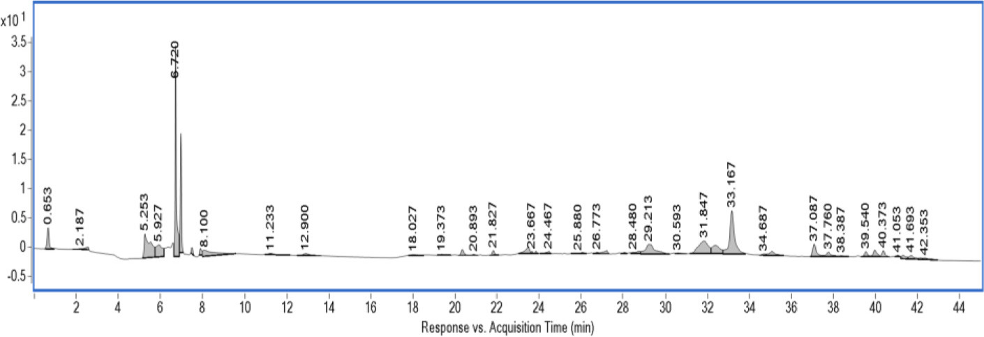
The methanolic extract of M. jalapa was subjected to LC-MS analysis to elucidate its phytochemical profile. The LC-MS chromatogram recorded at 254 nm revealed a diverse range of peaks, indicating the presence of multiple bioactive compounds. The results highlight a complex phytochemical matrix composed mainly of flavonoids, phenolic acids, and sterol-like compounds. Each peak represents a distinct compound, which was tentatively identified based on retention time (RT), observed molecular ion ([M+H]+), and comparison with theoretical values reported in literature and databases. The chromatogram displays a series of peaks distributed from 0.6 to 42 minutes. A very prominent and sharp peak was detected at RT 6.220 min, indicating the most abundant compound in the extract. Other moderate to minor peaks span across the chromatogram, with significant chemical diversity detected particularly between 5 and 35 minutes, where many flavonoid compounds are known to elute. The detection wavelength at 254 nm is sensitive to π→π* transitions, which are common in conjugated systems such as flavonoids, phenolics, and other aromatic plant metabolites. This wavelength was selected to maximize the detection of antioxidant and pharmacologically relevant compounds. Using the chromatographic data and matching the observed [M+H]+ values with published databases and literature, tentative identifications were made for each major peak. Table 2 summarizes the findings.
Table 1
Comparative HPTLC profiling of Mirabilis jalapa at different wavelength.
Table 2
Tentative identification of compounds in Mirabilis jalapa methanolic extract via LC-MS.
The LC-MS data of M. jalapa demonstrates the plant’s richness in phenolic and flavonoid compounds, many of which are well-documented for their medicinal and therapeutic applications. The most intense peak at RT 6.220 min corresponds to Rutin, a flavonoid glycoside that exhibits strong antioxidant, anti-inflammatory, and vasoprotective properties. Its high abundance indicates its potential role as a major bioactive compound in the extract.
Adjacent to this peak, at RT 5.927 min, another significant compound was identified as Quercetin-3-O-glucoside. This compound has been widely studied for its anticancer, antiviral, and cardioprotective actions. It is common in plant extracts and contributes significantly to antioxidant mechanisms by scavenging free radicals. Phenolic acids such as 4-Hydroxybenzoic acid (RT 2.187), Ferulic acid (RT 11.233), and Coumaric acid (RT 18.027) were also detected. These compounds are well known for their capacity to neutralize oxidative stress, reduce inflammation, and protect against microbial infections. Their presence supports the traditional use of M. jalapa in the treatment of wounds, infections, and inflammatory disorders. The presence of apigenin (RT 26.470), a flavone with anticancer and anti-inflammatory activity, and luteolin (RT 33.167), known for its neuroprotective and antioxidant potential, adds further value to the phytochemical composition. These compounds have been extensively researched for their role in modulating enzymatic pathways, including those related to cyclooxygenase and nitric oxide synthase.
Interestingly, β-sitosterol was observed at RT 30.953, a phytosterol involved in lowering cholesterol and modulating immune responses. This compound has demonstrated anti-inflammatory and anticancer potential in various studies and could play a role in the adaptogenic properties of M. jalapa. In the later part of the chromatogram, Kaempferol (RT 37.060) and Naringenin (RT 41.953) were detected. Both of these flavonoids are associated with antiaging, antidiabetic, and antimicrobial effects. Kaempferol, in particular, is recognized for its ability to modulate oxidative stress at the cellular level. All detected compounds were within ±5 ppm difference from their theoretical mass values, suggesting a high level of accuracy in mass spectral matching and high confidence in compound identification. Nevertheless, these identifications remain tentative and would benefit from further confirmation via MS/MS fragmentation data or co-elution with authentic standards.
The phytochemical profile revealed by LC-MS aligns well with the known pharmacological effects of M. jalapa. The diverse array of flavonoids and phenolics provides a biochemical rationale for the plant’s broad spectrum of biological activities. These compounds act in synergy, suggesting that the whole extract may offer more therapeutic benefit than isolated components. This supports the use of whole plant formulations in traditional medicine systems. The LC-MS analysis of M. jalapa methanolic extract has successfully revealed a wide range of bioactive constituents, predominantly flavonoids and phenolic acids. The presence of major compounds such as rutin, quercetin-3-O-glucoside, and ferulic acid supports the ethnomedicinal claims regarding this plant. The chromatographic fingerprint generated can serve as a chemical marker for standardization and quality control of M. jalapa-based formulations. Further detailed structural elucidation through tandem MS (MS/MS), nuclear magnetic resonance, and biological assays is necessary to fully characterize and validate the therapeutic roles of these phytochemicals.
3.5. Network pharmacology analysis
3.5.1. Compound and protein interaction
The network pharmacology interaction map constructed from the LC-MS analysis of M. jalapa reveals a comprehensive interaction between bioactive phytoconstituents and target genes involved in sexual dysfunction and aphrodisiac regulation. The network comprises multiple nodes representing bioactive compounds and their interacting gene targets, illustrating the multitarget therapeutic potential of the plant. Specifically, this interaction map highlights the involvement of at least 10 key phytochemicals, including quercetin, rutin, ferulic acid, coumaric acid, isoquercitrin, apigenin, luteolin, kaempferol, naringenin, and β-sitosterol. These compounds interact with more than 40 genes associated with various pathophysiological processes underlying sexual disorders, such as hormonal imbalance, oxidative stress, inflammation, neurodegeneration, and vascular dysfunction.
Among the compounds, quercetin emerges as a central hub with the highest degree of connectivity, indicating its wide-ranging pharmacological effects. Quercetin is known for its potent antioxidant, anti-inflammatory, and hormone-modulating properties. In the network, it shows direct interactions with key genes like TP53, APP, IL1B, IGF1, CYP19A1, STAT3, TNF, and BDNF. The gene TP53 plays a regulatory role in cell survival and apoptosis, and its modulation is essential in preserving reproductive tissue health under stress conditions. APP and BDNF are neurogenic factors, where BDNF in particular supports neural plasticity and sexual behavior regulation. IL1B and TNF are pro-inflammatory cytokines whose overexpression is often linked to erectile dysfunction and testicular damage. The interaction of quercetin with CYP19A1 (aromatase) implies its potential in regulating estrogen biosynthesis, vital for maintaining hormonal balance in both sexes. Similarly, its association with IGF1 points toward an influence on reproductive tissue development and spermatogenesis.
Figure 3
Thin layer chromatography (TLC) analysis of the methanolic extract of Mirabilis jalapa under different visualization conditions: (A) and (B) represent different samples or solvent systems. The first image shows the TLC plate under shortwave UV light (254 nm), revealing fluorescent spots. The second image displays the same under longwave UV light (366 nm), highlighting additional fluorescent compounds. The third image shows the plate after spraying with a derivatizing agent (e.g., vanillin-sulfuric acid) and heating, revealing colorless or colored spots. The Rf values and color intensities indicate the presence of multiple phytochemicals, including flavonoids, phenolics, and glycosides.

Figure 4
MS chromatogram of methanolic extract of Mirabilis jalapa recorded at 254 nm, showing multiple peaks at different retention times. Each peak corresponds to bioactive compounds potentially present in the extract. The chromatographic profile highlights the chemical diversity and complexity of the phytoconstituents within the sample.
Luteolin and apigenin also demonstrate substantial multitarget interactions with genes such as AR (androgen receptor), ABCA1, BDNF, CYP19A1, and TNF. Their capability to modulate AR and BDNF suggests roles in enhancing libido, neuroendocrine regulation, and mood stabilization. The AR is a pivotal receptor in male sexual physiology, responsible for mediating the effects of testosterone. A deficiency in androgen signaling is directly related to reduced sexual desire and erectile function. Thus, the positive interaction of flavonoids like apigenin and luteolin with AR reflects potential androgenic activity. Furthermore, apigenin’s link to ABCA1 and TNF suggests benefits in reducing oxidative stress and systemic inflammation, both of which impair reproductive function. Naringenin, another flavonoid depicted in the network, targets genes such as IGF1, AR, and ABCA1, adding to its role in promoting anabolic activity, testosterone modulation, and lipid homeostasis, which are crucial for sexual vitality.
The inclusion of β-sitosterol in the network is particularly relevant due to its structural similarity to cholesterol and known influence on androgen metabolism. It indirectly supports testosterone availability by inhibiting the conversion of testosterone to dihydrotestosterone, thereby potentially ameliorating benign prostatic hyperplasia and erectile dysfunction. The network indicates sitosterol interactions with a fewer number of genes compared to quercetin or apigenin, but these interactions (with key regulators like STAT3 and ESR1) suggest meaningful contributions to hormonal regulation and anti-inflammatory mechanisms. Kaempferol, isoquercitrin, and rutin also show interactions with genes including ESR1, APOE, and HSPB1. ESR1, the estrogen receptor gene, is crucial for sexual function in both males and females. Its modulation is essential for libido, lubrication, and vascular integrity. APOE is involved in lipid metabolism and indirectly affects testosterone levels, while HSPB1 has protective roles in stress responses within reproductive organs.
The genes interacting with multiple compounds, STAT3, IL6, and TNF, appear as central regulators within the inflammation and stress response axes. IL6 and TNF are commonly elevated in cases of erectile dysfunction, testicular inflammation, and psychosexual stress. Their downregulation or modulation via phytoconstituents like quercetin and apigenin may result in improved testicular function and sexual behavior. STAT3 is a transcription factor involved in immune and neuroendocrine responses. Its regulation influences prolactin and dopamine pathways, which are integral to sexual arousal and ejaculation control. Notably, EGFR and PIK3CA in the network suggest effects on reproductive cell proliferation and vascular smooth muscle relaxation, necessary for proper erectile function. These genes contribute to downstream signaling in nitric oxide pathways and vascular integrity critical in male sexual performance.
Further network analysis demonstrates that the bioactive compounds from M. jalapa does not operate via isolated targets but instead engages with overlapping and interconnected pathways that coalesce around key regulatory hubs. For example, the simultaneous modulation of TP53, IGF1, and BDNF by multiple compounds suggests a synchronized enhancement of neuroprotection, stress resilience, and reproductive cell viability. This polypharmacological approach mirrors traditional medicine’s holistic view, where one herb addresses multiple facets of a disorder. The redundancy and convergence observed in the network improve the robustness of therapeutic outcomes, particularly in multifactorial conditions like sexual dysfunction.
Moreover, the network offers insight into gender-specific influences. Genes such as CYP19A1, ESR1, and AR cater to both male and female sexual physiology. CYP19A1 is pivotal for estrogen synthesis, ESR1 governs estrogen receptor sensitivity, and AR modulates androgen response. In females, estrogen is vital for vaginal lubrication, sexual receptivity, and overall mood, whereas in males, androgens drive libido, sperm production, and erectile function. The compounds interacting with these genes (e.g., kaempferol, apigenin, and ferulic acid) suggest dual-gender efficacy, highlighting M. jalapa’s utility in broader sexual health applications.
The intensity of interaction—indicated by edge color and thickness further suggests the strength of each compound’s regulatory capacity. For example, the dense pink edges linking quercetin and apigenin with TNF, IL6, and STAT3 emphasize a probable potent anti-inflammatory action. This is highly relevant given that inflammation is a primary driver in several sexual disorders, including prostatitis, erectile dysfunction, and hypogonadism. Additionally, nodes such as BDNF and IGF1 represent neurotrophic and growth-promoting mechanisms that are not only essential for brain health but are also critical for sexual desire and performance. The presence of nodes like APP and APOE underscores potential links between cognitive health and sexual behavior, particularly in aging populations where dementia and decreased sexual interest often co-occur.
Moreover, the network pharmacological analysis of compounds derived from M. jalapa reveals an extensive interaction landscape with genes that are fundamentally involved in sexual dysfunction pathophysiology. This multicompound, multitarget framework demonstrates how traditional aphrodisiac herbs can exert broad-spectrum therapeutic effects through modern molecular pathways. The findings affirm that phytoconstituents such as quercetin, apigenin, and luteolin can significantly influence hormonal, neural, and vascular pathways via interaction with key regulators like AR, TP53, BDNF, IL6, and STAT3. These interactions support the traditional use of M. jalapa in managing sexual health and justify further exploration for targeted drug development or integrative therapeutic approaches in sexual medicine.
Figure 5
Network pharmacology interaction map illustrating the complex interplay between bioactive compounds identified from M. jalapa through LC-MS analysis and their predicted protein targets associated with sexual dysfunction. Nodes represent compounds (colored shapes) and target genes (colored circles), while edges denote molecular interactions. Key bioactive compounds such as quercetin, apigenin, luteolin, and sitosterol demonstrated strong multitarget binding, interacting with major genes like TP53, IL6, TNF, STAT3, and IGF1. This network reveals the polypharmacological potential of M. jalapa in modulating diverse molecular pathways linked to aphrodisiac activity and reproductive health, supporting its ethnomedicinal relevance and therapeutic promise.
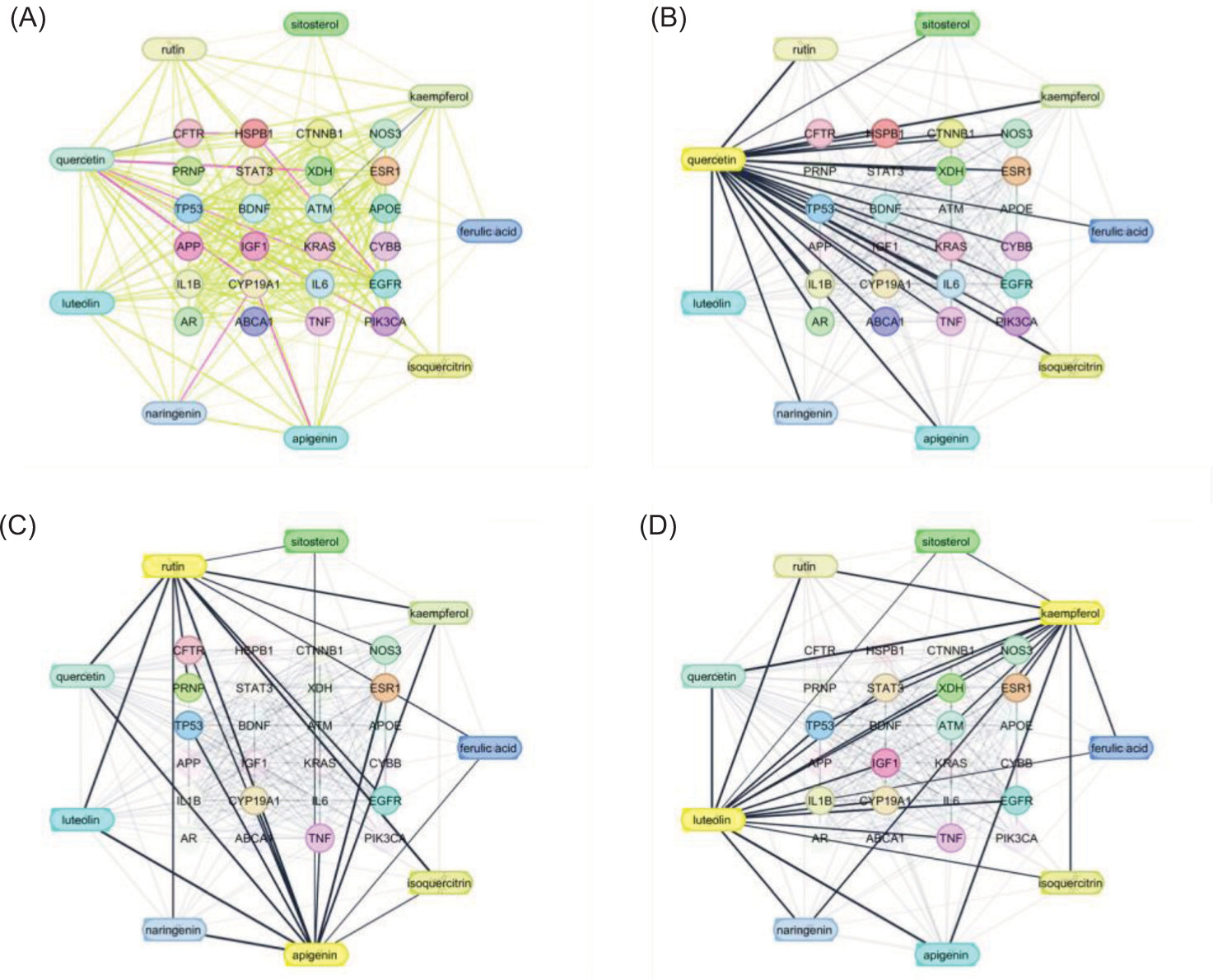
Table 3
List of the interacted genes along with their abbreviations, full names, and bioactive compounds from Mirabilis jalapa that showed interaction with each gene.
3.5.2. Gene ontology and DisGeNET analysis
The interpretation of GO and DisGeNET analysis provides valuable insights into the molecular mechanisms and disease associations related to sexual dysfunction. By integrating pathway and disease enrichment data, as illustrated in the provided image, one can elucidate the multifactorial basis of sexual disorders. This analytical approach bridges the gap between molecular signaling events and clinically observed pathologies, offering mechanistic explanations for how certain genes influence sexual health. The GO and pathway enrichment analysis using −log10(p) values, which indicate the significance of gene enrichment across various biological processes and pathways. The most significant pathway identified is Urotensin II-mediated signaling (WP5158), a vasoconstrictor peptide known to influence cardiovascular function and smooth muscle activity. This is highly relevant to erectile function, which relies on proper vascular tone and endothelial health. Dysregulation of such pathways may impair penile blood flow, thereby contributing to erectile dysfunction. Furthermore, the regulation of miRNA transcription (GO:1902893) highlights the importance of epigenetic control mechanisms in sexual health, as miRNAs are known to regulate gene expression posttranscriptionally, affecting hormone levels, receptor sensitivity, and vascular homeostasis.
The regulation of phosphorylation (GO:0042325) and cell activation (GO:0001775) also emerge as enriched processes. Phosphorylation is a key signaling mechanism in cellular responses to hormonal stimuli, particularly testosterone and estrogen, which modulate libido and reproductive organ function. These processes are crucial in neuronal signaling, vascular reactivity, and even spermatogenesis, suggesting that defects in phosphorylation cascades may manifest in various forms of sexual dysfunction. Negative regulation of cell population proliferation (GO:0008285) and apoptotic signaling (GO:2001233) indicate that abnormal cell turnover, particularly in reproductive or vascular tissues, may contribute to degenerative changes or tissue fibrosis associated with sexual disorders. The Integrated breast cancer pathway (WP1984) and proteoglycans in cancer (hsa05205) might seem tangential, but they highlight the shared molecular machinery between sexual dysfunction and hormone-dependent cancers such as breast and prostate cancer. These overlaps can inform therapeutic repurposing or warn of side effects of cancer treatments on sexual function. Similarly, pathways like regulation of smooth muscle cell proliferation (GO:0048660) are directly implicated in erectile function, given the role of smooth muscle in corpus cavernosum physiology. Inhibition or excessive proliferation in these cells can lead to fibrosis or penile structural changes, impairing erectile response. Additional insights come from cellular responses to steroid hormone compounds (GO:1901699) and lipid metabolism (GO:0071396), which are foundational to hormone synthesis and signaling. Disorders in these pathways can lead to low testosterone or estrogen levels, disrupting libido, erectile function, and vaginal lubrication. Moreover, genes involved in VEGFA-VEGFR2 signaling (R-HSA-4420097) are key to angiogenesis and vascular integrity. Dysfunction in these pathways could cause microvascular complications, a known contributor to erectile dysfunction, especially in diabetic patients (Gaurav et al., 2022).
Figure 6
Gene ontology and DisGeNet analysis of Mirabilis jalapa L. metabolites in aphrodisiac activity and via regulation of different pathways.
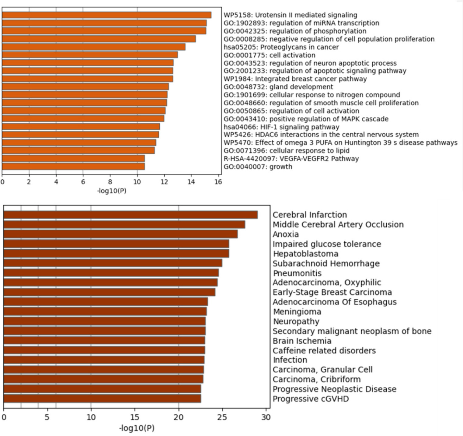
In the DisGeNET analysis, which correlates genes with specific diseases based on statistical significance. The top associations include cerebral infarction, middle cerebral artery occlusion, and anoxia, all of which are neurological or vascular conditions (Jiang et al., 2020; Yusuf et al., 2021). These results suggest a strong neurovascular component to sexual dysfunction, reinforcing the notion that healthy brain function and adequate blood supply are essential for sexual arousal and performance. For instance, cerebral infarction may impair areas of the brain involved in sexual desire or autonomic regulation of genital blood flow. Further diseases like impaired glucose tolerance and neuropathy are closely linked to diabetes, a well-known risk factor for sexual dysfunction. Hyperglycemia can damage nerves (diabetic neuropathy) and blood vessels, both of which are critical to erectile function and genital sensation. Similarly, subarachnoid hemorrhage, pneumonitis, and hepatoblastoma suggest systemic health impacts that may indirectly contribute to sexual dysfunction through hormonal imbalance, psychological distress, or medication side effects (Marumo et al., 2001; Park et al., 2021; Yu et al., 2021).
Moreover, the presence of various carcinomas (e.g., adenocarcinoma, early-stage breast carcinoma, granular cell carcinoma) in the analysis suggests a possible link between genes involved in oncogenesis and sexual health. This could be due to hormonal dependencies in these cancers, where therapeutic suppression of sex hormones leads to decreased libido and sexual capability. Furthermore, secondary malignant neoplasm of bone and brain ischemia add to the complexity, indicating that widespread systemic illness or central nervous system injury can severely impact sexual functioning. Diseases such as caffeine-related disorders and Progressive cGVHD (chronic graft-versus-host disease) indicate lifestyle or immune system interactions with sexual health. Caffeine, while a common stimulant, has controversial effects on sexual performance, potentially impacting blood flow or anxiety levels. Graft-versus-host disease often requires immunosuppressive therapy, which can reduce hormonal levels or interfere with reproductive organ integrity (Asiaf et al., 2014; Farias et al., 2021; Filippone et al., 2023).
This dual analysis paints a comprehensive picture: sexual dysfunction is not an isolated problem but a manifestation of disruptions in neurovascular, endocrine, and cellular processes. It also shows that sexual dysfunction shares genetic underpinnings with a broad spectrum of diseases, including metabolic, cardiovascular, neurological, and oncologic conditions. This overlap underscores the importance of a holistic approach in diagnosis and treatment—one that goes beyond mere symptomatic relief and addresses the root molecular causes. Moreover, understanding the exact role of these genes and their pathways opens new avenues for targeted pharmacological interventions and personalized medicine strategies. Network biology thus becomes a powerful tool in unraveling the multifaceted etiology of sexual dysfunction and improving outcomes through precision therapeutics.
4. CONCLUSION
The present investigation comprehensively elucidates the phytochemical composition and aphrodisiac potential of M. jalapa L. root extract through LC-MS analyses and network pharmacology. Chromatographic analyses revealed a chemically diverse profile with classes of compounds previously implicated in reproductive and hormonal modulation. Network pharmacology mapped these metabolites to key molecular targets and pathways associated with sexual function, including androgen receptor regulation, nitric oxide biosynthesis, and steroid hormone biosynthesis. Moreover, protein-metabolite interaction analysis identified significant associations with genes such as TP53, APP, IL1B, IGF1, CYP19A1, STAT3, and TNF, which play crucial roles in reproductive, vascular, and neuroendocrine regulation. Protein-protein interaction networks and GO enrichment analyses further substantiated these findings. Collectively, the study provides robust scientific validation for the traditional use of M. jalapa root as an aphrodisiac and establishes a strong foundation for in vitro, in vivo, and clinical investigations toward developing safe, efficacious phytotherapeutic agents for sexual dysfunction.