

Introduction
In December of 2019, the world was struck by a new type of pneumonia with symptoms varying from fever to dry cough and dyspnea. The emergence of the virus causing this disease took place in Wuhan, China. Initially, that illness was named Wuhan pneumonia since the number of cases was restricted to the Wuhan region. However, due to the rapid virus spread and its associated disease’s high infection rate in other countries, the World Health Organization (WHO) considered this pathogen a global threat and declared its resulting disease a pandemic on March 11, 2020. Through sequencing of the virus genome, it was possible to uncover that the causative agent of this newly discovered pneumonia belongs to the coronavirus family, which is knowingly capable of infecting humans (Liu, Kuo, & Shih, 2020).
Coronaviruses are RNA-type microorganisms widely spread among humans, other mammals and birds, and they can cause respiratory, enteric and neurological disorders. The etiological agent of the aforementioned atypical pneumonia has been named Severe Acute Respiratory Syndrome Coronavirus 2 (SARS-CoV-2), and its consequent disease has been named Coronavirus Disease 2019 (COVID-19). Initially, the SARS-CoV-2 spike glycoprotein binds to human angiotensin-converting enzyme 2 (ACE2) in order to infect cells. Then, its transcription machinery produces four structural proteins, namely envelope small membrane (E), spike (S), nucleocapsid (N) and membrane (M), but also a myriad of accessory proteins, such as open reading frame (ORF) 3a, 8 and 9b, as well as replicase polyproteins that are proteolytically cleaved into sixteen nonstructural proteins (Nsps), including papain-like protease/PLpro (Nsp3), 3C-like protease (Nsp5), Nsp9, RNA-directed RNA polymerase/RdRp (Nsp12) and helicase (Nsp13) (Liu et al., 2020).
After the pathophysiology of SARS-CoV-2 was discovered, an incessant search began for a drug or compound capable of fighting its replication and consequent infectious disease process. Different drugs and active ingredients have been tested at the same time that several countries have started a race to produce an effective vaccine against COVID-19 (Wu et al., 2020). Numerous steps are necessary to verify both the efficacy and the safety of potential new drugs when it comes to validating studies involving SARS-CoV-2. Techniques pertaining to areas of Molecular Biology, Bioinformatics, Immunology, Pharmacodynamics and Pharmacokinetics are imperative for the development of compounds that can fight COVID-19 (Pan et al., 2013). In this context, molecular docking is a computational experiment that aims to search for the leading interactions between a target (receptor/protein) and a given ligand. This bioinformatic technique takes into account the best fit as well as the finest interaction comprising a target, e.g., protein binding sites, and the specific geometry of a ligand, so that an associated binding energy score can be generated. In this type of experiment, specific in silico techniques are used to analyze protein-ligand interactions as well as different ligand conformations that are obtained as a result of a complex formed with a protein’s active or allosteric site (Azevedo & Walter, 2019).
Nowadays, one of the most widely used computer programs on the molecular docking realm is AutoDock Vina, a software developed by (Trott & Olson, 2010) and whose main application is to perform rigid-flexible molecular docking screenings. In this type of in silico methodology, the chosen target remains rigid (with no rotation, no translation and no torsion), while its associated ligand has enough flexibility to generate distinct conformations (positions), out of which a favorable interaction can be established (Trott et al., 2010).

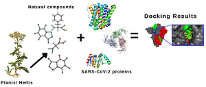
Medicinal plants have been utilized to combat a multitude of pathogens. This important fact may be due to the existence of various bioactive compounds found in natural plants (Figure 1). As a result, a great variety of drug classes originated from natural plants has emerged. Teas, poultices, decoctions and a myriad of other different forms of both extracting herbs’ medicinal constituents and administering them have been used for centuries. Additionally, in the last decades, evaluations have been conducted to establish their promising applications on a therapeutic scope (Atanasov et al., 2015).
Natural compounds have a remarkable advantage over synthetic substances. Although both of these chemical entities have the same structure and therefore equal physicochemical and biological properties, compounds obtained from medicinal plants do not need to be synthetized. As a positive outcome, they do less harm to the environment since extraction techniques may use smaller quantities of organic solvents, which are, for the most part, knowingly toxic (Fuzimoto & Isidoro, 2020). For this reason, we analyzed several natural chemical compounds with the aim of verifying their efficacy against SARS-CoV-2 proteins through in silico investigations.
Material and Methods
All natural compounds that comprise the Food and Drug Administration (FDA) library were chosen as ligands and then downloaded. Subsequently, all selected FDA structures were converted to *.pdbqt format using AutoDock Tools (ADT) v1.5.6, which was used as a graphical interface (GUI) to edit and generate all converted files.
With regard to the quest for potential targets in SARS-CoV-2, the majority of proteins that we chose participate in the initial infection as well as in the subsequent viral replication process that characterizes COVID-19. A total of 24 proteins were chosen from PDB (Protein Data Bank) website (https://www.rcsb.org), and the most adequate files (the ones exhibiting the highest resolution) were downloaded in *.pdb format, which contains all information relating to the elected structures. Proteins were also prepared using ADT GUI. Each 3D protein structure file in *.pdb format was separately inserted and read into ADT GUI. Then, polar hydrogen atoms were added, and water molecules as well as other unwanted ligand molecules were removed so that there was no bias due to the presence of these chemical entities on the binding sites. Finally, all files were saved in *.pdbqt format.
Table 1
General characteristics of all SARS-CoV-2 proteins selected for molecular docking studies.
Molecular docking assays were performed using AutoDock Vina v1.2.3. To ensure that ligands would interact with the regions where they exhibit the best fit in each of the 24 chosen proteins, molecular docking calculations were delimitated by a 3D space called grid box, in which Vina’s search algorithms test different positions with an eye to find the first-rate ligand conformation that complexes with the given target. The aforementioned grid box delimitations encompassed the whole proteins, for we wanted to extend our search to cover potential allosteric sites.
Results for all proteins and their respective complexed ligands were arranged in an increasing energy order (from most negative to most positive) for the purpose of determining the finest interaction profiles. Then, once the most favorable results had been obtained, the best ranked conformations of every ligand associated with each macromolecule were analyzed in order to dissect interaction profiles (Figure 2). In this context, we used PyMOL v2.5 as a molecular visualization system.

We analyzed all results from a scoring function perspective, also called score, which performs variation in rotational, translational, and conformational positions as well as in motions to find the most stable target-ligand complex. Aside from that, hydrogen bonds (H-bonds) were also determined. To this end, we utilized Maestro v13.0. Finally, to validate our in silico results, we conducted redocking, and Root-Mean-Square Deviation (RMSD) values were calculated using AutoDock Tools (ADT) v1.5.7 (Figure 3).

Results and Discussion
In an attempt to find leading drugs and bioactive substances to either prevent or treat COVID-19, we noticed that green tea contains catechins and polyphenols, both of which may be effective in combating this disease. Additionally, these chemical entities are promising drug candidates capable of impairing viral replication, and their activities against different types of viruses have inspired us to search for auspicious compounds capable of inhibiting SARS-CoV-2 initial infection and/or replication (Calland et al., 2012; Carneiro, Batista, Braga, Nogueira, & Rahal, 2016; Ismail & Jusoh, 2017).
Research studies show that several ligands are able to interact with various SARS-CoV-2 proteins, and their results exhibit a first-rate binding affinity between a given ligand and its associated protein (Senger et al., 2020). This type of binding energy between two molecules is based on the "lock and key" scheme, a well-known strategy in computational drug design. The protein-ligand or even protein-protein perfect fit is mainly based on this concept. Specific interactions between two structures occur through H-bonds, van der Waals forces as well as other interacting bonds that result in a complex between two given molecules. In this context, researchers look for the lowest possible energy to generate stability, which takes place when two substances that form a complex come together, as previously described by the "lock and key" scheme (Chen, Seukep, & Guo, 2020). Molecular docking screenings using AutoDock Vina v1.2.3 enabled us to find first-rate positions defined by binding energy scores between given ligands and their respective SARS-CoV-2 macromolecules (Table 1).
Table 2
Descriptions of all SARS-CoV-2 proteins selected for molecular docking studies.
Various bioactive substances, well-known for treating both neuroinflammation and neurodegenerative diseases, have demonstrated great interaction profiles with SARS-CoV-2 proteins. Punicalagin is a natural compound present in pomegranate (Punica granatum) and consumed worldwide. This fruit is used in popular medicine because it helps treat chronic diseases, such as diabetes, atherosclerosis and inflammatory processes (Akhtar, Ismail, Fraternale, & Sestili, 2015; Banihani, Swedan, & Alguraan, 2013; Danesi & Ferguson, 2017; Igdoura et al., 2013). The majority of bioactive compounds found in pomegranate, namely phytobiotics, flavonoids and anthocyanins, are from its peel. These substances possess therapeutic properties, including anti-inflammatory, antioxidant, hypoglycemic and antimicrobial effects (Grabež et al., 2020; Lansky & Newman, 2007; Saeed et al., 2018; Xu et al., 2022).
Table 3
Molecular Docking Results of Bioactive Natural Compounds against SARS-CoV-2 proteins.
As showed in Table 2, punicalagin interacted with several macromolecules, mostly proteins related to SARS-CoV-2 replication process, such as RdRp-nsp7-nsp8 complex (7C2K, 7BV1 and 7BV2), 3CLpro, also named main protease – Mpro (7C2Q and 6LU7, in both immature and mature states, respectively), PLpro (6WX4), Nsp-16-Nsp10 complex proteins (6W4H and 6WKS), ORF3a protein (6XDC), Nsp7-Nsp8 replicase complex (6XIP) and Nsp-1 protein (7K3N). In addition, punicalagin also interacted with spike glycoproteins (6LXT and 6ZGE). The best energy score results are the ones from the punicalagin-RdRp-nsp7-nsp8 complex. The interaction profile between punicalagin and 6ZGE is characterized by four H-bonds established with amino acid residues PRO 2222, HIS 2232, SER 3115 and SER 3123, all of which are disclosed in the scheme that represents (Figure 4).

Solamargine is a substance found in tomatoes (Solanum lycopersicum), potatoes (Solanum tuberosum), aubergine/eggplant (Solanum melongena) as well as in other plants belonging to Solanum genus species. That natural compound was described by (Tang et al., 2022) as an anticancer substance that can effectively treat various types of malignant tumors, such as hepatocellular carcinoma (HCC). Its alpha form is a knowingly bioactive alkaloid found in Solanum surattense, which previously showed great binding affinity with SARS-CoV-2 Mpro (Hasan et al., 2020). Our results, on the other hand, exhibit that alpha-solamargine interacted through better binding energy scores with a spike-ACE2 complex (6M0J) as well as with PLpro macrodomain (6WEY). H-bond interactions between solamargine and 6M0J are shown on Figure 5.
Licorice (Glycyrrhiza glabra) is a plant whose roots’ chemical substances have been used as antioxidants to prevent cancer as well as natural compounds to inhibit the growth of Leishmania major promastigotes and amastigotes in infected mice. Additionally, these chemical entities have been utilized to treat bacterial, fungal and viral infections (Chrzanowski, Chrzanowska, & Graboń, 2020; Sheikhi, Khamesipour, Radjabian, Ghazanfari, & Mohammadi, 2022). In 2005, Hoever and collaborators discovered through molecular docking studies that glycyrrhizinate dipotassium and ammonium glycyrrhizinate, bioactive molecules found in Glycyrrhiza glabra, could increase antiviral activity of glycyrrhizin derivatives against SARS-CoV (Hoever et al., 2005). In 2020, (Chrzanowski et al., 2020) disclosed that the aforementioned licorice chemical compounds enhanced glycyrrhizin potential binding to ACE2 (Chrzanowski et al., 2020). Our results show a probable multitarget function of licorice as glycyrrhizinate dipotassium and ammonium glycyrrhizinate interacted through extremely favorable binding energy scores of -10.7 kcal/mol and -10.5 kcal/mol with SARS-CoV-2 helicase/Nsp13 (5RL9) and nucleoprotein (6ZCO), respectively. In regard to 5RL9, glycyrrhizinate dipotassium formed three H-bonds with amino acid residues VAL 752, THR 770 and THR 974. All binding interactions established between glycyrrhizinate dipotassium and 5RL9 as well as between ammonium glycyrrhizinate and 6ZCO can be seen in detail on Figure 7; Figure 6, respectively.


Saikosaponin A (SSA) A, saikosaponin B2 (SSB2) and saikosaponin D (SSD) are compounds present in sickle-leaved hare's-ear (Bupleurum falcatum), an endemic European and Western Asian plant that has anti-ulcer properties as well as the capacity to inhibit the growth of colon tumor cells in vitro (Sun, Matsumoto, & Yamada, 1991; Zhang et al., 2022). Our molecular docking studies evince that SSA, SSB2 and SSD favorably interacted with SARS-CoV-2 ORF9b protein (6Z4U), envelope small membrane protein (7K3G) and PLpro (6W9C), respectively, exhibiting favorable binding energy scores of -8.2 kcal/mol, -8.9 kcal/mol and -9.7 kcal/mol, one by one. All H-bond interactions established between SSA and 6Z4U, SSB2 and 7K3G as well as between SSD and 6W9C can be visualized in detail on Figure 10; Figure 9; Figure 8, respectively.
Notoginsenoside R1 and asiaticoside are bioactive compounds present in two traditional Chinese medicinal herbs, namely Panax notoginseng and Gotu Kola (Centella asiatica), respectively. Immune stimulation effects (Zhang et al., 2016) and alleviation of chronic hypoxic pulmonary hypertension are two Panax notoginseng therapeutic properties in mice (Chen, Zeng, & Lu, 2017; Wang et al., 2015). Pertaining to SARS-CoV-2, our results show that notoginsenoside R1 favorably interacted with Nsp9 replicase (6WXD), having exhibited two H-bonds with amino acid residues ARG 10 and ARG 221 (Figure 11). On the other hand, Gotu Kola is commonly used to improve scarring and to treat skin injuries, such as lupus, ulcers and psoriasis, as well as to help combat diarrhea and fever (Chen et al., 2017). In regard to the new coronavirus, asiaticoside exhibited a great binding interaction with spike glycoprotein S1 subunit (7C01). All H-bonds established between notoginsenoside R1 and 6WXD as well as between asiaticoside and 7C01 can be seen in detail on Figure 12; Figure 11, respectively.
Only the ORF8 protein (7JTL) out of 24 SARS-CoV-2 macromolecules did not demonstrate a favorable binding affinity with any of all chemical substances tested in our molecular docking screenings. The remarkable energy score values found in our research study involving bioactive natural compounds may be explained by means of ligand-protein stabilization through hydrogen bonds observed from our in silico point of view.
Conclusions
In essence, all aforementioned binding interaction profiles disclosed by our molecular docking study reveal that bioactive natural compounds derived from plants and herbs can negatively affect SARS-CoV-2 infection process. The capacity of these chemical entities to interact with essential proteins of the new coronavirus reaffirm researchers’ perspective regarding the imperativeness of continuing to study phytochemicals against pathogens. Furthermore, natural compounds may be more environmentally friendly concerning the use of solvents, for there is no need of a synthetic reaction step when it comes to extracting chemical substances.
Hence, all results found in our research study indicate a valuable forthcoming application of promising bioactive natural compounds derived from plants and herbs, such as Punica granatum, Centella asiatica and Solanum spp., in the realm of both in vitro and in vivo studies that will corroborate with our findings against COVID-19.